|
||
|
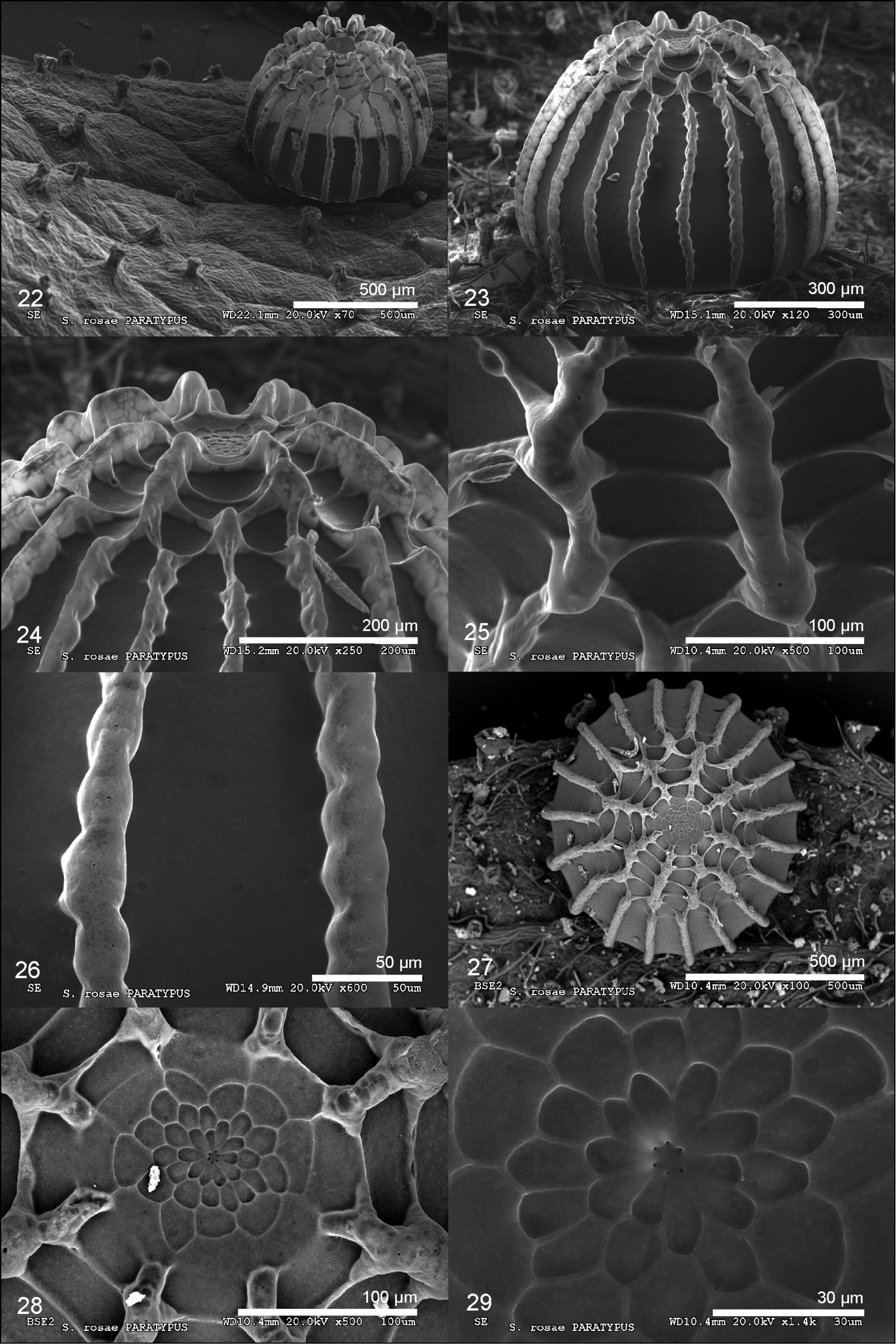
Scanning electron microscope images of the egg of Spialia rosae (Paratypes from Puerto de la Ragua, Granada, SE Spain). 22. Lateral view of egg on a leaf of Rosa sicula showing the glands of the plant epidermis. 23. Lateral view. 24. Lateral view of the annular area with annular area in the middle. 25. Cells from the lateral zone of the egg formed by radial and transverse ribs. 26. Detail of the radial ribs. 27. View of the annular pole. 28. Annular area with the micropylar rosette. 29. Detail of the micropylar rosette and the micropyle showing six micropylar openings. |