|
||
|
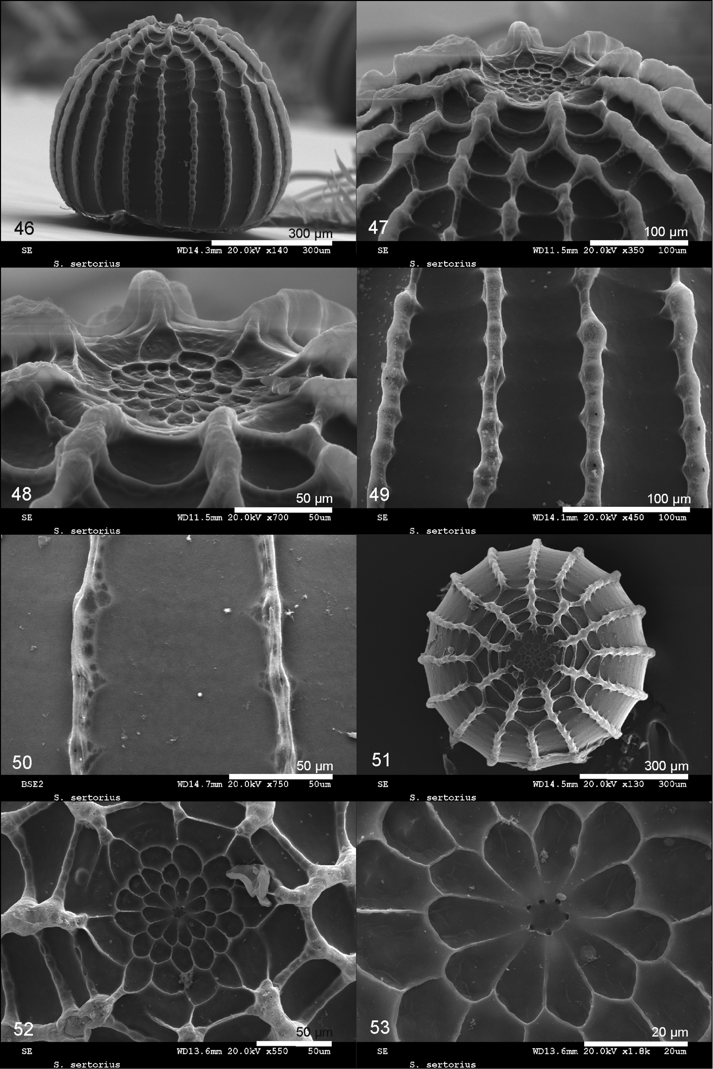
Scanning electron microscope images of an egg of Spialia sertorius from Vallelado, Segovia, Central Spain. 46. Lateral view. 47. Lateral view of the annular area. 48. Detail of the annular area. 49. Cells of the egg equator formed by radial and transverse ribs. 50. Detail of the radial ribs. 51. View of the annular pole. 52. Annular area with the micropylar rosette. 53. Micropylar rosette and micropyle showing six micropylar openings. |