|
||
|
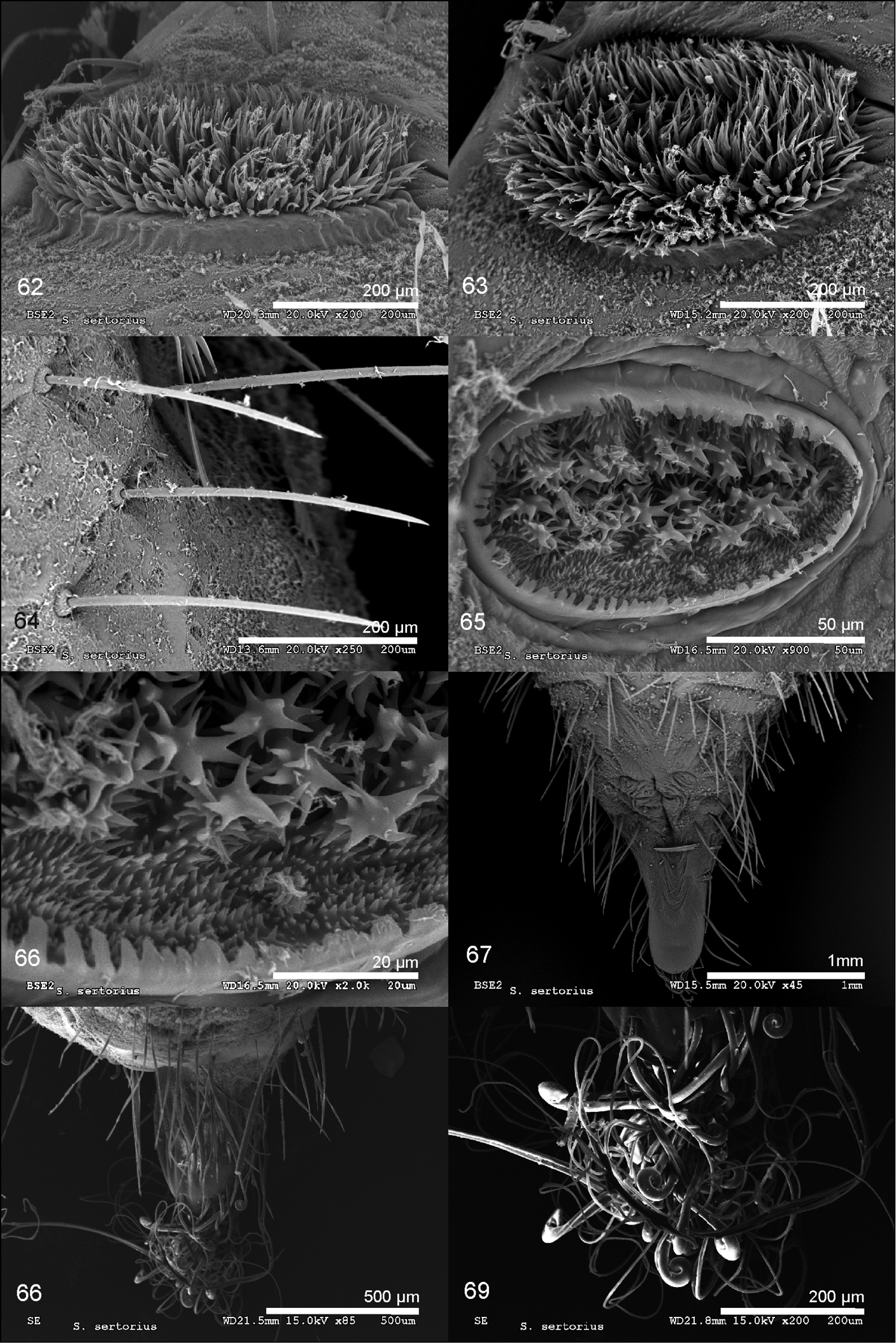
Scanning electron microscope images of a pupa of Spialia sertorius from Losar de la Vera, Cáceres, Central Spain. 62. Lateral view of the mesothoracic tubercle. 63. Upper view of the mesothoracic tubercle. 64. Detail of the pupal cuticle showing smooth setae with rounded bases. 65. Abdominal spiracle. 66. Detail of the spiny papillae inside the spiracle. 67. Ventral view of the last abdominal segments. 68. Detail of the cremaster 69. Detail of the cremastral hooks. |