|
||
|
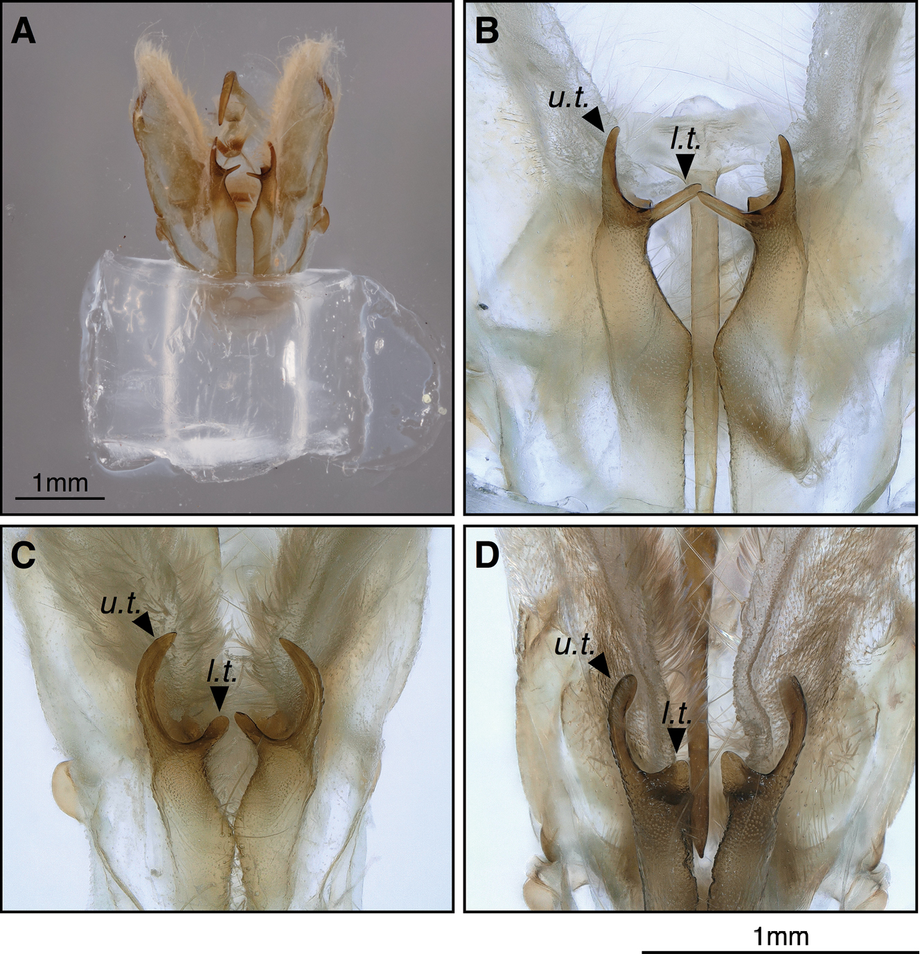
Unmounted genitalia capsule with closed valvae in ventral view in the tunnel-shaped holder. A. Overview of a T. sabaudiata genitalia placed in the tunnel; B–D: Close up photography of the sacculus projection, B. T. sabaudiata; C. T. dubitata and D. T. tauteli. Abbreviations: u.t. upper ‘thorn’ of sacculus projection; l.t. lower ‘thorn’ of sacculus projection. The sacculus projections appear diagnostic in this angle. |